Results Leukemia/lymphoma Panel By Flow
This panel would also be used for prognostication in these neoplasms. Additional FISH or molecular testing may be recommended by the.
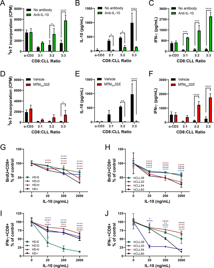
Interleukin 10 Suppression Enhances T Cell Antitumor Immunity And Responses To Checkpoint Blockade In Chronic Lymphocytic Leukemia Leukemia
Results showed that in 916 56 percent the diagnosis of lymphoma or cancer could be suspected by flow cytometry alone while 416 were consistent with the.

Results leukemia/lymphoma panel by flow. If possible submit CBC results with differential or an EDTA tube of peripheral blood. The proteins are markers that may help diagnose leukemia or lymphoma. B-cell leukemialymphoma panel is a blood test that looks for certain proteins on the surface of white blood cells called B-lymphocytes.
If no abnormalities are detected by the initial panel no further flow cytometric assessment will be performed unless otherwise indicated by specific features of the clinical presentation or prior laboratory results. Flow cytometric leukemia and lymphoma analysis may aid in identifying the tumor lineage for diagnostic and prognostic purposes. This test is usually done after abnormal results are seen on a complete blood count or WBC differential.
How the Test is Performed. Place 2 mL minimum volume. Immunophenotyping is a type of flow cytometry used to diagnose leukemia or lymphoma.
Flow Cytometry Leukemia and lymphoma analysis by flow cytometry aids in identifying the tumor lineage which in most cases is identified as T cell B cell or myeloid. The markers antigens that are present on the cells as detected by flow cytometry immunophenotyping will help characterize the cells present. With immunophenotyping your results will state whether any abnormal cells are present and what types of.
LeukemiaLymphoma Nonacute Marker Panel. Do not freeze and do not place in fixative. After review of the clinical history and morphology a panel of markers is selected for each case by a board-certified hematopathologist.
Flow cytometry - leukemialymphoma immunophenotyping B-cell leukemialymphoma panel is a blood test that looks for certain proteins on the surface of white blood cells called B-lymphocytes. Lineage identification can provide a confirmatory diagnosis or differential diagnosis prognosis and treatment options. 1 mL of bone marrow in a green top sodium heparin tube.
Flow cytometry can identify the type of cells in a blood or bone marrow sample including the types of cancer cells. Please complete a Request for Flow Cytometry Testing form and forward it with the specimen. If no abnormalities are detected by the initial panel no further flow cytometric assessment will be performed unless otherwise indicated by specific features of the clinical presentation or prior laboratory results.
Sometimes flow cytometry helps to define the leukemia subgroup. In normal or reactive processes a bimodal distribution of - and -positive B cells is present in a ratio of approximately 151. For example the so called M7 the megakaryoblastic leukemia.
LeukemiaLymphoma Combo Marker Panel. Diffuse Large B-Cell Lymphoma DLBCL LeukemiaLymphoma Acute Marker Panel. The proteins are markers that may help diagnose leukemia or lymphoma.
These tests may suggest lymphoma or leukemia but more information is generally needed to confirm a diagnosis and to identify a specific type of leukemia or lymphoma. Because of the critical nature of these specimens the laboratory will attempt to. If no abnormalities are detected by the initial panel no further flow cytometric assessment will be performed unless otherwise indicated by specific features of the clinical presentation or prior laboratory results.
Immunophenotyping using multiparameter analysis simultaneous staining with a pan B-cell marker and anti-immunoglobulin light chain antibodies is a rapid and specific method for detecting and confirming the presence of neoplastic B-cell disorders. The flow cytometric analysis of a specimen includes the use of a selected panel of leukocyte-associated antibodies to determine the lineage of a malignant or abnormal population of leukocytes. FlowLeukemia PanelFlow Cytometry LeukemiaLymphomaLymphoma PanelLeukemiaLymphoma Phenotyping PanelImmunophenotyping.
Label the specimen appropriately blood. Additional FISH or molecular testing may be recommended by the. Analysis is based on a CD45 gating strategy.
How the Test is Performed A blood sample is needed. Results are reported in percentages and relative intensities and are evaluated by a pathologist. We recommend this more limited panel for lymphoid neoplasms lymphoma lymphocytic leukemia or when a lymphoid origin is suspected for a leukemia based on morphologic features of the cells in Wrights-stained smears.
Plasma Cell Multiple Myeloma. Prolymphocytic Leukemia PLL Mantle Cell LymphomaLeukemia MCL Hairy Cell Leukemia HCL Non-Hodgkins Lymphoma. B lymphocyte cell surface markers.
Indicate blood on the request form. A laboratory report will typically include specific results from the tests as well as an analysis of what those results mean. Flow cytometry usually can give the answer.
Additional FISH or molecular testing may be recommended by the. Flow cytometry immunophenotyping may be performed on blood bone marrow or other samples to provide this additional information. Green top sodium heparin tube.
It detects types of cancer cells based on either the presence or the absence of certain protein markers antigens on a cells surface. Flow cytometry also helps to define lymphoma subgroups for example the distinction between CLL hairy cell leukemia.
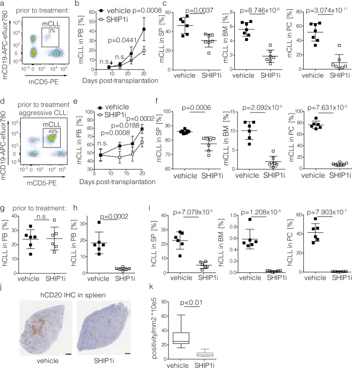
Targeted Pi3k Akt Hyperactivation Induces Cell Death In Chronic Lymphocytic Leukemia Nature Communications

Results Of Immunocytochemical Reactions In Acute Leukemia Download Table

A Real World Perspective Of Cd123 Expression In Acute Leukemia As Promising Biomarker To Predict Treatment Outcome In B All And Aml Clinical Lymphoma Myeloma And Leukemia
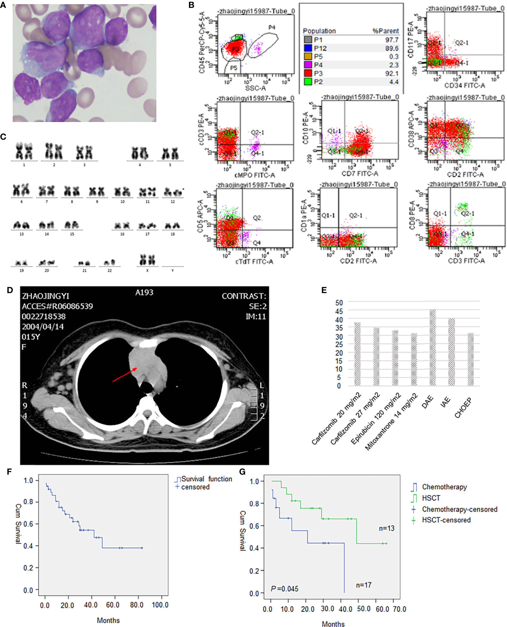
Frontiers Determining The Appropriate Treatment For T Cell Acute Lymphoblastic Leukemia With Set Can Nup214 Fusion Perspectives From A Case Report And Literature Review Oncology

Target Values For Markers Used In Cll Mrd Analysis Download Table
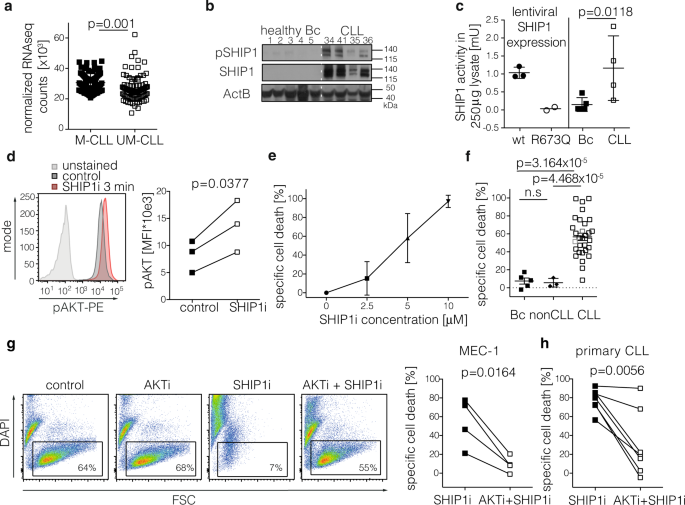
Targeted Pi3k Akt Hyperactivation Induces Cell Death In Chronic Lymphocytic Leukemia Nature Communications
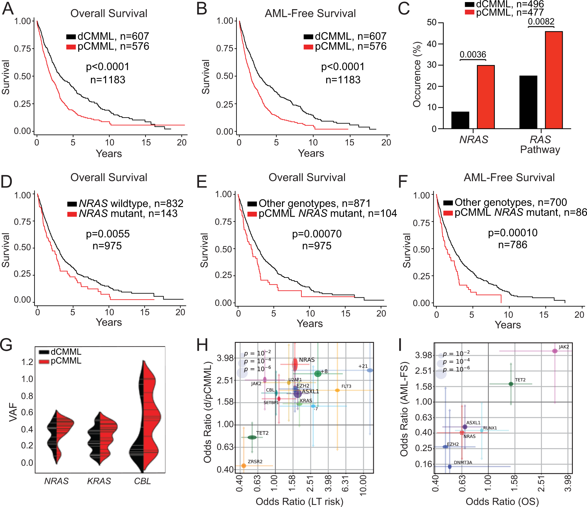
Ras Mutations Drive Proliferative Chronic Myelomonocytic Leukemia Via A Kmt2a Plk1 Axis Nature Communications
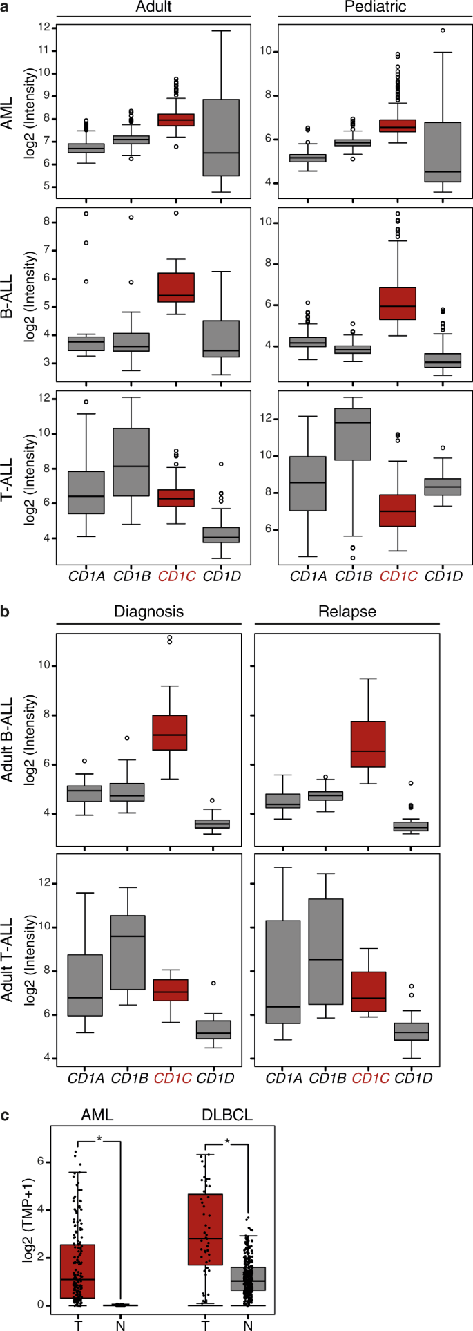
Human T Cells Engineered With A Leukemia Lipid Specific Tcr Enables Donor Unrestricted Recognition Of Cd1c Expressing Leukemia Nature Communications

Arv 825 A Brd4 Inhibitor Leads To Sustained Degradation Of Brd4 With Broad Activity Against Acute Myeloid Leukemia And Overcomes Stroma Mediated Resistance By Modulating Chemokine Receptor Cell Adhesion And Metabolic Targets

Pdf The Use Of The Antibodies In The Diagnosis Of Leukemia And Lymphoma By Flow Cytometry

Morphological Appearances Of Chronic Lymphocytic Leukaemia Cll And Download Scientific Diagram

Deletion Of Genes Encoding Pu 1 And Spi B Leads To B Cell Acute Lymphoblastic Leukemia Associated With Driver Mutations In Janus Kinases Biorxiv
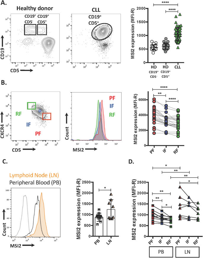
Musashi 2 Influences Chronic Lymphocytic Leukemia Cell Survival And Growth Making It A Potential Therapeutic Target Leukemia

Peripheral Blood Chronic Lymphocytic Leukemia Minimal Residual Disease Download Scientific Diagram

Mnda Controls The Expression Of Mcl 1 And Bcl 2 In Chronic Lymphocytic Leukemia Cells Experimental Hematology
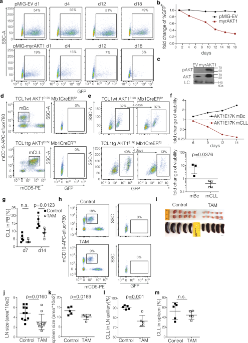
Targeted Pi3k Akt Hyperactivation Induces Cell Death In Chronic Lymphocytic Leukemia Nature Communications

Image Result For Leukemia Vs Lymphoma Hodgkins Disease Lymphoma Leukemia

Target Values For Markers Used In Cll Mrd Analysis Download Table
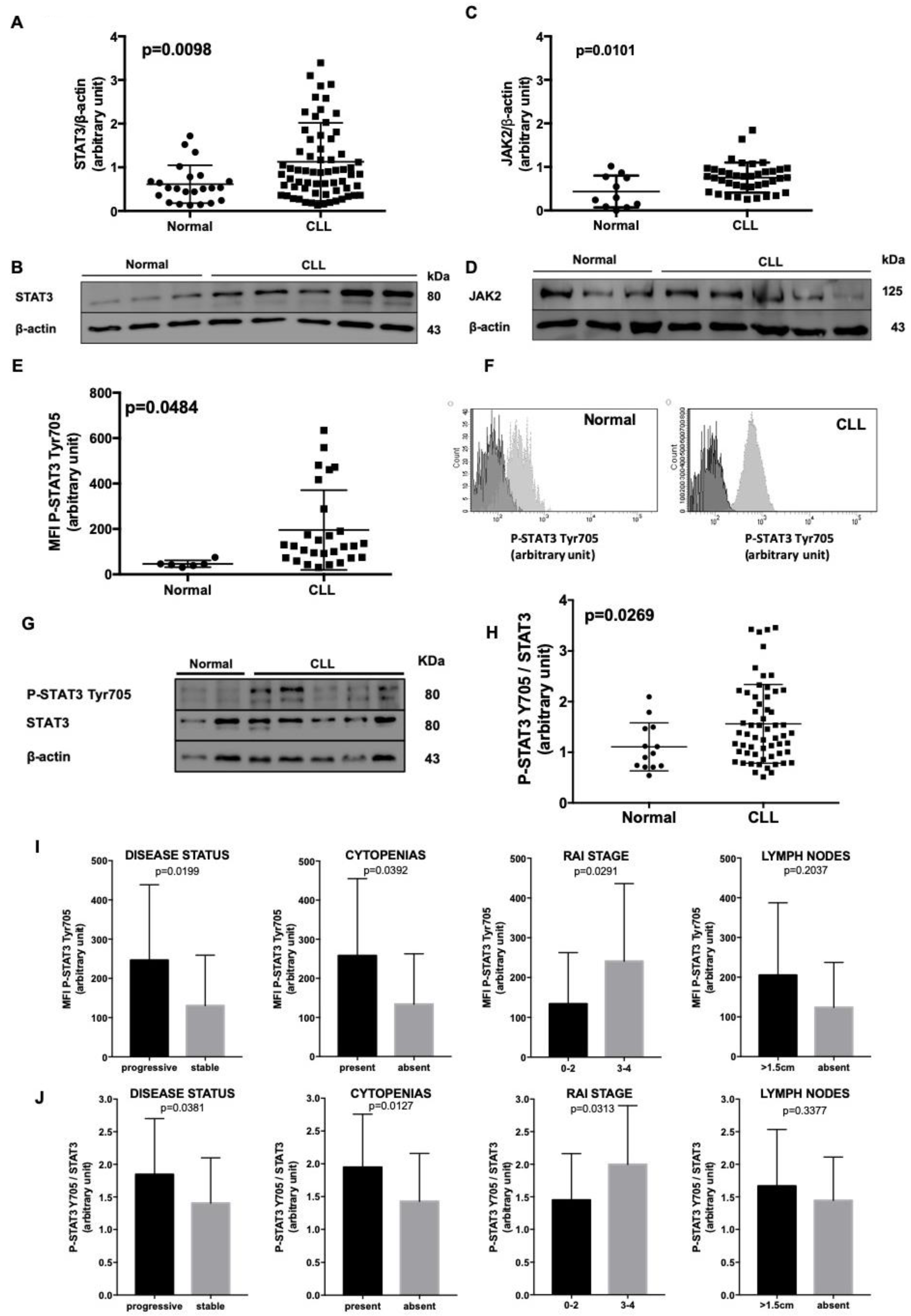
Cancers Free Full Text In Chronic Lymphocytic Leukemia The Jak2 Stat3 Pathway Is Constitutively Activated And Its Inhibition Leads To Cll Cell Death Unaffected By The Protective Bone Marrow Microenvironment Html
Posting Komentar untuk "Results Leukemia/lymphoma Panel By Flow"